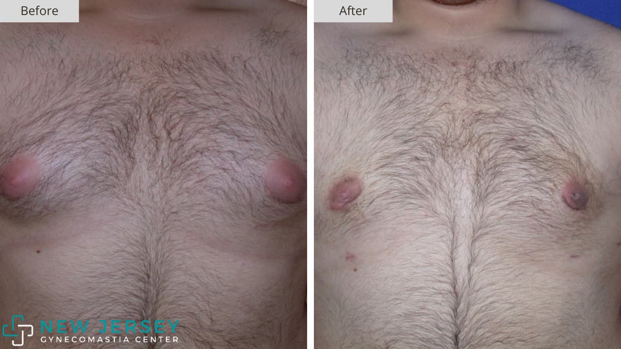
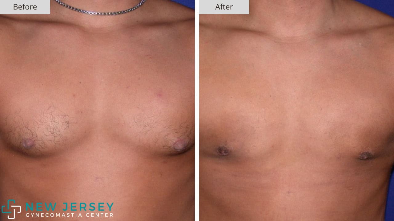
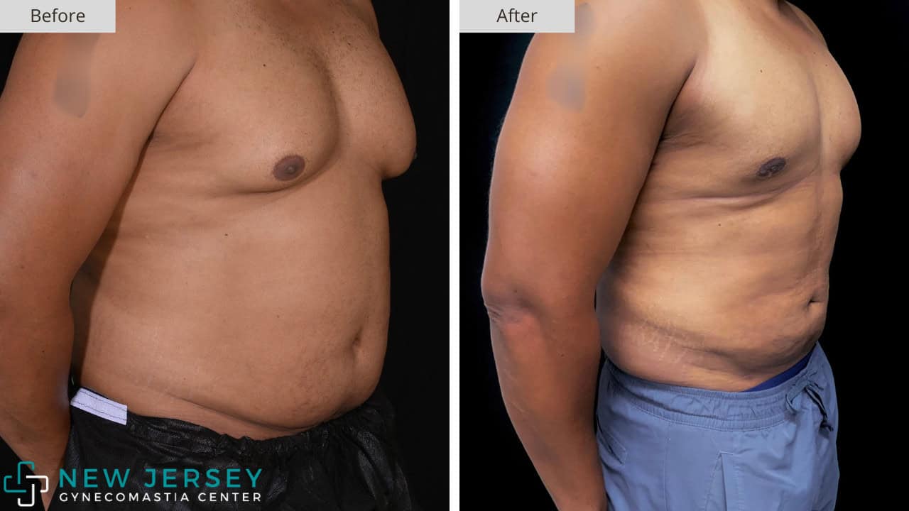
Are you wondering what your chest may look like after having gynecomastia surgery treatment in NJ?
Take a look at the photo collection below that shows before and after results for those who opted to have surgery with our doctors. This can also give you a better sense if you are a candidate for gyno surgery.
Feel free to learn more about the various stages of gynecomastia to understand where you fall on the spectrum, and take a look at what some of our former patients have had to say about their experiences.
Patient #1-A
- Grade: Grade 1
- Body Type: Average Build
This patient presented with raised, puffy areolas and some chest fullness. The glandular tissue was concentrated directly beneath each nipple, which created the visible projection seen in the before image. Treatment involved gland excision through a small periareolar incision on each side. The after photo shows nipples that now sit flat against the chest wall.
Patient #1-B
- Grade: Grade 1
- Body Type: Average Build
This lean patient had isolated nipple puffiness on an otherwise flat chest. The condition was purely glandular, and there was no excess fat that needed to be addressed. A small periareolar incision allowed for full removal of the firm tissue beneath each areola. The result is a smooth chest contour that matches his overall build.
Patient #1-C
- Grade: Grade 1
- Body Type: Average Build
This patient had localized glandular tissue under both areolas with no lateral fullness across the chest. Skin tone was a key consideration when the surgical plan was developed. The incision was placed along the lower areolar border so that the scar would stay concealed within the natural pigment line. The after image shows almost no visible evidence of the procedure.
Patient #1-D
- Grade: Grade 1
- Body Type: Average Build
Glandular tissue formed two firm mounds beneath the areolas on this patient. The surrounding chest fat was minimal, so liposuction was not part of the procedure. Direct excision through a small areolar incision removed the gland on each side. The chest now reads as flat and even with no remaining puffiness.
Patient #1-E
- Grade: Grade 1
- Body Type: Average Build
This patient showed visible areolar stretching in the pre-op photo, which was caused by glandular pressure from below. The condition was limited to the breast tissue itself, and there was no broader chest involvement. Gland excision was performed with careful attention to the areolar contour. The areolas now appear smaller and sit flush with the surface of the chest.
Patient #1-F
- Grade: Grade 1
- Body Type: Average Build
This was a subtle case with mild glandular projection at the nipples. The patient had no surrounding fat and no skin laxity to address. A hidden incision was placed at the lower areolar border to allow for full gland removal. The after photo shows a flat chest with very minimal scarring.
Patient #1-G
- Grade: Grade 1
- Body Type: Average Build
The oblique view of this patient revealed nipple projection that was less obvious from the frontal angle. The glandular tissue was concentrated directly under the areola, and there was no lateral component to address. Targeted excision flattened the area without any disruption to the surrounding pec contour. The side profile now shows an even line across the chest.
Patient #1-H
- Grade: Grade 1
- Body Type: Athletic Build
This patient had a built, athletic chest with strong pec development, but puffy nipples still showed through and threw off the shape. Even with low body fat and serious training, the gland tissue under each areola would not respond to diet or workouts. The procedure focused only on gland removal through small periareolar incisions.
Patient #2-A
- Grade: Grade 2
- Body Type: Average Build
Both sides of this chest had clear breast tissue that gave it a rounded shape. The areolas looked stretched from the gland sitting underneath. Surgery removed the gland along with a small amount of fat. The after photo shows an even chest with no leftover bulge.
Patient #2-B
- Grade: Grade 2
- Body Type: Average Build
A medium amount of breast tissue sat on each side of this patient's chest. The soft, round shape did not match his lean upper body. Both gland and fat were removed to bring the chest in line with his frame. His result is a flatter chest with a more typical male shape.
Patient #2-C
- Grade: Grade 2
- Body Type: Athletic Build
This patient had a heavily built chest with deep pec definition, but raised nipples and a soft fold under each pec gave away the gynecomastia. To match the rest of his physique, the procedure removed the gland and used light liposuction to clear the area without taking away from his hard-earned shape. The after photo shows a chest with the same muscle definition, only now flat and clean across the nipple line.
Patient #2-D
- Grade: Grade 2
- Body Type: Average Build
Large, dark areolas were the first thing visible in the before photo, and soft breast tissue underneath gave each side a round, full look. Fat removal and gland excision worked together to flatten the chest and reduce the pressure pushing the areolas outward. As a result, the areolas now sit flat against the surface of the chest.
Patient #2-E
- Grade: Grade 2
- Body Type: Average Build
The before image shows a cone-shaped chest with full, raised nipples, which happens when gland pushes the areola forward and up. The procedure focused on gland removal with some light fat work to smooth out the surrounding area. With the gland gone, the chest now has a smooth, even surface across the front.
Patient #2-F
- Grade: Grade 2
- Body Type: Athletic Build
This patient came in with soft, rounded chest tissue on both sides, and the nipples sat puffy and full against the rest of his chest hair. Liposuction handled the fat while gland excision took out the firm tissue underneath, which gave a complete result. The after photo shows a chest that looks more athletic and balanced with the rest of his upper body.
Patient #2-H
- Grade: Grade 2
- Body Type: Larger Build
A heavier build can make Stage 2 gynecomastia harder to spot, but the chest tissue is still there and shows up as a soft round shape. This patient had a mix of gland and fat, so surgery cleared out the gland and used liposuction to slim the chest area at the same time. The after photo shows a flatter chest that fits the rest of his body without the rounded look.
Patient #2-I
- Grade: Grade 2
- Body Type: Larger Build
The frontal view shows breast fullness on both sides of the chest, but a side angle makes the issue more clear with a visible bulge above the pec line. To address both layers of tissue, the procedure used liposuction along with gland removal for a complete result. The side view after surgery shows a flat profile from shoulder to belly with no remaining fullness.
Patient #2-J
- Grade: Grade 2
- Body Type: Larger Build
Side views often tell the real story with Stage 2 gynecomastia, and this patient's chest pushed forward in a clear cone shape with the nipple sitting at the top. Gland removal flattened the chest and softened the slope, which made a major difference from this angle. The after side view now shows a natural, even line with no projection at the nipple.
Patient #2-K
- Grade: Grade 2
- Body Type: Average Build
Even fullness on both sides was the main concern for this patient, and the angled view in the second set shows how far the chest stuck out at the nipple. Both gland and fat were addressed during surgery to get a complete result from every angle. The after photos show a flat chest with a clearer pec shape, both from the front and from the side.
Patient #2-L
- Grade: Grade 2
- Body Type: Larger Build
A heavier upper body can make chest tissue blend in, but the puffiness still shows, especially in the side photo where the raised, full nipple stands out. To address the issue, liposuction handled the fat while gland excision cleaned up the firm tissue underneath. The frontal after photo shows a chest that fits better with his overall shape and looks more natural in every view.
Patient #3-A
- Grade: Grade 3
- Body Type: Larger Build
The before photo shows two distinct breast mounds with a clear fold line at the bottom, which is a hallmark of Stage 3. Both gland and fat tissue had built up, so the procedure addressed both layers along with some skin tightening at the surface. The after photo shows a flat chest where the fold line is gone and the nipples sit in a more masculine position.
Patient #3-B
- Grade: Grade 3
- Body Type: Larger Build
This patient had clear breast tissue on each side with stretched skin that had lost some of its tone. To handle the full picture, the procedure combined gland excision, liposuction, and skin tightening to flatten the chest in one session. The after image shows a smooth, even surface with no leftover fold or sagging.
Patient #3-C
- Grade: Grade 3
- Body Type: Larger Build
The before photos from both the front and the side show full breast shape with the areolas pointing slightly downward. Surgery removed the gland, took out fat with liposuction, and tightened the skin so it would lie flat against the new contour. The result is a chest that looks tight and athletic, with strong pec definition visible from every angle.
Patient #3-D
- Grade: Grade 3
- Body Type: Larger Build
Stretched skin and a visible breast crease made this a clear Stage 3 case from the start. Both gland and fat were removed during the procedure, and the skin was tightened so it could settle smoothly over the new shape. The after photo shows a flat chest with the crease fully gone and the nipples in a more natural spot.
Patient #4-A
- Grade: Grade 4
- Body Type: Larger Build
This patient had heavy breast tissue with skin that had stretched and lost its tone over time. The nipples sat lower on the chest than they should, which is common with Stage 4 cases. Surgery removed the gland and fat while also tightening the skin so it could lay flat against the new chest shape.
Patient #4-B
- Grade: Grade 4
- Body Type: Larger Build
The before photo shows full breast shape with clear sagging and downward-pointing nipples, which marks this as a Stage 4 case. To get a complete result, the procedure combined gland removal, liposuction, and major skin tightening in one session. The after image shows a flat chest with the nipples lifted into a more masculine spot and no leftover sag.
Patient #4-C
- Grade: Grade 4
- Body Type: Larger Build
This patient came in with heavy breast tissue and stretched skin that had pulled the areolas downward and outward. The procedure removed the gland and fat, and the areolas were lifted internally to keep them from drooping after surgery.
Note: Sunken appearance expected to resolve as recovery progresses due to internal suspension of areola to avoid drooping.
At the New Jersey Gynecomastia Center, we’re all about giving you top-notch care from start to finish. From your very first consultation to your recovery and beyond, we’re here for you every step of the way. We’re honored to be entrusted with your care, and we take that responsibility seriously. Our team, led by board-certified gynecomastia plastic surgeon specialists, is dedicated to providing the absolute best in patient care and outcomes. We understand that every person is unique, so we tailor our approach to suit your individual needs. Got questions? Ready to schedule your in-person or virtual consultation? Don’t hesitate to reach out to the New Jersey Gynecomastia Center today. We’re here to help!
New Jersey Gynecomastia Center
1567 Palisade Avenue #3A
Fort Lee, New Jersey 07024
(551) 201-1110
